Product Manager: Harrison Michael
Cardiovascular diseases, which pose a significant threat to global health, include a variety of conditions such as hyperlipidemia and atherosclerosis, hypertension, myocardial infarction, heart failure, arrhythmias, and shock. Animal models are crucial tools for in-depth exploration of the pathogenesis of these diseases and for evaluating therapeutic interventions. This article will systematically and comprehensively describe the methods for establishing various animal models of cardiovascular diseases and the corresponding experimental protocols, providing a comprehensive reference for relevant scientific research.
Selection of Common Experimental Animals
There are significant differences among various experimental animals in terms of cardiovascular system structure, physiological function, and susceptibility to diseases. The rational selection of experimental animals is the primary prerequisite for successful model establishment.
1.Rats: With strong reproductive capacity, low breeding costs, and convenient handling, rats are widely used in the construction of models such as hypertension and myocardial infarction. For example, spontaneously hypertensive rats (SHR) are commonly used for the study of primary hypertension.
2.Mice: With mature gene-editing technology, mice can be used to construct transgenic or gene knockout models, which are suitable for exploring the mechanisms of diseases such as atherosclerosis and heart failure. The ApoE gene knockout mouse is a classic model for atherosclerosis research.
3.Rabbits: Sensitive to cholesterol metabolism, rabbits are prone to forming atherosclerotic plaques after consuming high-cholesterol feed, making them suitable for observing early lesions of atherosclerosis.
4.Miniature pigs: With a cardiovascular system highly similar to that of humans, miniature pigs are particularly suitable for preclinical studies such as interventional therapy and medical device development. However, they have high breeding costs and greater operational complexity.
5.Dogs: With cardiac anatomical structure and physiological functions similar to those of humans, dogs are often used in the construction of models such as arrhythmias and shock, facilitating electrocardiographic monitoring and hemodynamic studies.。
Methods and Experimental Protocols for Establishing Animal Models of Cardiovascular Diseases
(I) Animal Models of Hyperlipidemia and Atherosclerosis
1. Diet-induced method
◦ Experimental animals: Rabbits, mice, rats
◦ Experimental protocol
▪Rabbit model: Feed a high-fat diet containing 1% cholesterol and 5% lard to the animals for 8 - 12 weeks. Regularly monitor body weight and blood lipid levels on a weekly basis, and dynamically observe the morphological changes of the arterial vessels through ultrasound.
▪Mouse model: Use ApoE gene knockout mice or LDLR gene knockout mice, and feed them a Western diet (containing 21% fat and 0.15% cholesterol) for 12 - 16 weeks. Collect blood at certain intervals to detect key blood lipid indicators such as total cholesterol (TC), triglycerides (TG), and low-density lipoprotein cholesterol (LDL-C).
◦ Precautions: Strictly control the nutritional component ratios of the feed to avoid animal death due to overfeeding; closely monitor blood lipid indicators to ensure successful model establishment.
2. Balloon injury method
◦ Experimental animals: Rabbits, miniature pigs
◦ Experimental protocol
▪After anesthesia, insert a balloon catheter through the femoral artery or carotid artery to the target artery (such as the abdominal aorta).
▪Inflate the balloon to a pressure of 6 - 8 atm and slowly pull it back and forth 3 - 5 times to cause endothelial injury to the blood vessel.
▪Postoperatively, feed the animals with regular feed and raise them for 4 - 8 weeks, observing plaque formation through angiography and histological staining.
◦ Precautions: Strictly follow aseptic principles during the operation to prevent complications such as blood vessel rupture and infection; accurately control the balloon pressure and pulling times to avoid excessive vascular injury.
(II) Animal Models of Hypertension
1. Renal hypertension model (two-kidney, one-clip method)
◦ Experimental animals: Rats
◦ Experimental protocol
▪After anesthesia, expose one kidney, separate the renal artery, and use a 0.2 - 0.25mm silver clip to clamp the main trunk of the renal artery while preserving the other kidney.
▪Postoperatively, regularly measure the tail arterial blood pressure using the tail-cuff method. When the systolic blood pressure remains above 160mmHg, the model is considered successful.
◦ Precautions: The surgical operation should be gentle to avoid damaging the kidney tissue; select a silver clip of the appropriate size to prevent incomplete or excessive clamping that may lead to kidney necrosis.
2. Spontaneous hypertension model
◦ Experimental animals: Spontaneously hypertensive rats (SHR)
◦ Experimental protocol
▪Raise SHR in a standard environment with free access to food and water, measure blood pressure weekly, and meticulously record the trend of blood pressure changes. Blood pressure-lowering drugs can be administered to observe therapeutic effects.
◦ Precautions: Strictly control the breeding environment temperature (22±2℃) and humidity (50±10%) to minimize the impact of environmental factors on blood pressure.
(III) Animal Models of Myocardial Infarction
1. Coronary artery ligation method
◦ Experimental animals: Rats, mice, miniature pigs
◦ Experimental protocol
▪Perform tracheal intubation on the animals and connect them to a ventilator for assisted breathing.
▪Open the chest to expose the heart, and ligate the left anterior descending branch of the left coronary artery 1 - 2mm below the lower edge of the left atrial appendage using 6 - 0 or 8 - 0 silk thread.
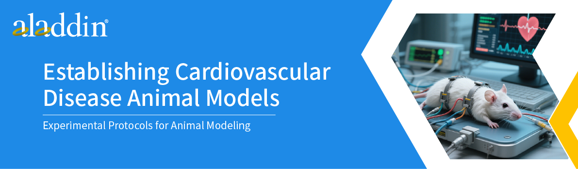
▪Postoperatively, administer antibiotics to prevent infection, monitor ST-segment elevation in real-time through electrocardiogram (ECG), and assess cardiac function using echocardiography.
◦ Precautions: Accurately control the depth of anesthesia to avoid respiratory depression due to excessive depth or animal restlessness due to insufficient depth; ensure accurate ligation position to guarantee a stable myocardial infarction area.
2. Drug-induced method
◦ Experimental animals: Rats
◦ Experimental protocol
▪Intraperitoneally inject isoproterenol (ISO) at a dose of 5 - 10mg/kg, once daily for 3 - 7 consecutive days. Assess the degree of myocardial damage through myocardial enzyme spectrum detection (such as creatine kinase isoenzyme CK-MB, lactate dehydrogenase LDH) and cardiac tissue pathological analysis.
◦ Precautions: Strictly control the drug dosage and injection frequency to prevent animal death due to drug toxicity.
(IV) Animal Models of Heart Failure
1. Rapid ventricular pacing method
◦ Experimental animals: Dogs, miniature pigs
◦ Experimental protocol
▪Insert a pacing electrode through the jugular vein or femoral vein into the right ventricle, and pace the ventricle at a frequency of 200 - 400 beats per minute for 2 - 4 weeks.
▪Regularly detect key cardiac function indicators such as left ventricular ejection fraction (LVEF) and left ventricular end-diastolic diameter (LVEDD) through echocardiography.
◦ Precautions:Ensure accurate electrode positioning to prevent displacement; closely monitor the pacing frequency to avoid arrhythmias or animal death due to excessively high frequency.
2. Doxorubicin-induced method
◦ Experimental animals: Rats, mice
◦ Experimental protocol
▪Administer doxorubicin through tail vein injection, with a total dose of 15 - 20mg/kg, divided into 3 - 5 injections, each separated by 3 - 5 days. Observe the animal's activity status and weight changes, and assess the degree of heart failure through cardiac function detection.
◦ Precautions: Doxorubicin has cardiotoxicity and bone marrow suppression as side effects. Closely monitor the animal's condition and promptly manage adverse reactions.
(V) Animal Models of Arrhythmias
1. Drug-induced method
◦ Experimental animals: Rats, mice, dogs
◦ Experimental protocol
▪Rats/Mice: Intraperitoneally inject aconitine at a dose of 10 - 15μg/kg to rapidly induce arrhythmias such as ventricular premature beats and ventricular tachycardia.
▪Dogs: Intravenously inject epinephrine at a dose of 0.01 - 0.1mg/kg to induce rapid arrhythmias; or intravenously inject verapamil at a dose of 0.1 - 0.2mg/kg to induce slow arrhythmias.
▪After drug injection, immediately monitor electrocardiographic activity continuously through ECG and record parameters such as the type and duration of arrhythmias.
◦ Precautions: Ensure accurate electrode positioning to prevent displacement; closely monitor the pacing frequency to avoid arrhythmias or animal death due to excessively high frequency.
2. Electrical stimulation method
◦ Experimental animals: Dogs, miniature pigs
◦ Experimental protocol
▪After anesthesia, puncture percutaneously or open the chest to expose the heart and fix the electrodes on the surface of the atrium or ventricle.
▪Apply electrical stimulation of different frequencies, intensities, and durations, such as using short bursts of rapid pacing with a frequency of 100 - 500 beats per minute and a stimulation duration of 5 - 10 seconds, which can induce atrial fibrillation or ventricular fibrillation.
▪Record electrocardiographic changes in real-time through a multi-channel ECG to assess the success of the arrhythmia model.
◦ Precautions: Accurately fix the electrode position to avoid affecting the stimulation effect; strictly control the electrical stimulation parameters to prevent irreversible cardiac damage due to excessive stimulation.
(VI) Animal Models of Shock
1. Hemorrhagic shock model
◦ Experimental animals: Rats, rabbits, dogs
◦ Experimental protocol
▪After anesthesia, expose the femoral artery or carotid artery and insert an arterial catheter.
▪Bleed at a certain rate to reduce the animal's mean arterial pressure (MAP) to 40 - 50mm Hg and maintain this blood pressure level for 1 - 2 hours. The bleeding rate should be adjusted according to the animal's body weight, generally controlled at 1 - 2ml/min/kg.
▪Continuously monitor hemodynamic indicators such as MAP, heart rate (HR), and central venous pressure (CVP), as well as biochemical indicators such as blood gas analysis during the bleeding process.
◦ Precautions: Strictly control the amount and rate of bleeding to avoid animal death due to excessive and rapid blood loss; keep the animal warm during the experiment to prevent hypothermia from affecting the experimental results.
2. Endotoxin shock model
◦ Experimental animals: Rats, mice
◦ Experimental protocol
▪Induce shock through intravenous injection of lipopolysaccharide (LPS). The injection dose for rats is 5 - 10mg/kg, and for mice, it is 1 - 5mg/kg. After injection, observe the animal's mental state and temperature changes, and detect the levels of inflammatory factors (such as TNF-α, IL-6) and hemodynamic indicators to assess the degree of shock.
◦ Precautions: The LPS injection dose should be optimized through pre-experiments based on the animal species and strain; closely monitor the animal's condition during the experiment and promptly manage severe adverse reactions.
Model Evaluation and Validation
1.Physiological index detection: Regularly measure basic physiological indicators such as animal blood pressure, heart rate, respiratory rate, body temperature, and body weight, and observe changes in animal behavior and mental state.
2.Hematological detection: Collect blood to detect biochemical indicators such as blood lipids, myocardial enzyme spectrum, brain natriuretic peptide (BNP), inflammatory factors, and coagulation function to assess disease progression and systemic response.
3.Imaging detection: Utilize techniques such as echocardiography, magnetic resonance imaging (MRI), computed tomography (CT), and angiography to observe changes in cardiac structure and function, as well as vascular lesions.
4.Histological analysis: After euthanizing the animals, take relevant tissues such as the heart, blood vessels, and kidneys for pathological sectioning. Observe tissue morphological changes and pathological characteristics through methods such as HE staining, Oil Red O staining, and Masson staining.
5.Molecular biology detection: Use techniques such as PCR and Western blot to detect the expression levels of related genes and proteins, and deeply explore the molecular mechanisms of disease occurrence and development.
Aladdin:https://www.aladdinsci.com/