Licia Miller Product Manager
Platelet separation is a technique for separating and enriching platelets from whole blood by physical or chemical methods. It has important applications in medical and biological research. For example, separated platelets can be used for platelet function research, signal pathway analysis, disease marker detection, etc. In clinical practice, platelet-rich plasma (PRP) can also be prepared for tissue repair and regenerative medicine.
To prevent platelet activation during manipulation, strong mechanical forces (e.g., rapid pipetting, vigorous shaking) should be avoided. In addition, researchers may choose to add inhibitors that are appropriate for their research and experimental goals. All buffers during the experiment should be stored and used at 4°C unless otherwise stated.
Blood collection precautions: (1) It is important to ensure that potential donors have not taken medications that could interfere with platelet studies and platelet function (e.g., antihistamines, aspirin, nonsteroidal anti-inflammatory drugs) in the two weeks prior to blood draw. (2) Polypropylene, polyethylene, or polycarbonate labware is recommended for handling and storing platelets. (3) Blood is a body fluid and should be considered a biohazard. Protective gear should always be worn, and blood should be handled according to the researcher's institution's regulations.
Phase 1 Blood fractionation
First, whole blood is obtained and platelet-rich plasma (PRP) is prepared by centrifugation. Centrifugation conditions can vary widely (e.g., from 800 × g for 5 minutes to 100 × g for 20 minutes), but good separation results are always obtained.
The phlebotomist should draw blood into a Becton Dicinson Vacutainer® (with ACD, yellow cap) or a plastic syringe containing 1/10 volume of CPD buffer. The amount of blood required depends on the experiment. In general, 40 - 45 mL of blood will yield an average of 1 - 3 × 10⁹ platelets.
The required buffer reagents and formulas are as follows:
(1) ACD buffer (acid-citrate-dextrose)
39 mM citric acid, 75 mM sodium citrate, 135 mM glucose, pH 7.4.
Before use, warm to room temperature and adjust pH as needed.
(2) CPD buffer (citrate-phosphate-dextrose)
16 mM citric acid, 90 mM sodium citrate, 16 mM NaH2PO4, 142 mM glucose, pH 7.4.
Before use, warm to room temperature and adjust pH as needed.
Experimental Steps
1. The blood collector shall collect blood according to the strict blood collection process. After collection, the blood shall be placed at 18-25°C immediately without shaking and processed within 2 hours.
Centrifuge at 200 × g for 20 minutes at room temperature.
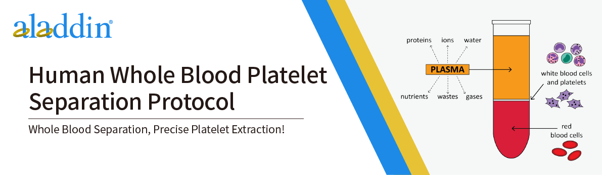
3. After centrifugation, the blood is separated into layers:
Lower layer: Red blood cells (50 - 80% of the total volume)
Intermediate layer (grey-white layer): A very thin band of white blood cells (also called the "buffy coat")
Upper layer (yellow): PRP (platelet-rich plasma)
4. Use a wide-mouth pipette to gently aspirate the upper layer of PRP into a new tube (avoid aspirating the middle layer of white blood cells).
Phase II Platelet separation
Experimental Steps
1.Add HEP buffer to PRP at a 1:1 ratio (v/v). Add prostaglandin E1 (PGE1, 1 µM final concentration) to prevent platelet activation.
HEP buffer (HEP refers to HEPES in the buffer) formula:140 mM NaCl, 2.7 mM KCl, 3.8 mM HEPES, 5 mM EGTA, pH 7.4.
Before use, warm to room temperature and adjust pH as needed.
2. Slowly invert the test tube and mix gently.
3. Spin at 100 × g for 15 - 20 minutes at room temperature (without deceleration) to pellet contaminating red and white blood cells.
4. Use a wide-bore pipette to transfer the supernatant to a new plastic tube.
5. Centrifuge at 800 × g for 15 - 20 minutes at room temperature (without stopping the machine) to pellet the platelets and discard the supernatant.
6. Rinse the platelets by gently adding wash buffer and then slowly removing it with a pipette (no need to resuspend to avoid unnecessary platelet activation).
Platelet washing buffer formula:10 mM sodium citrate, 150 mM NaCl, 1 mM EDTA, 1% (w/v) glucose, pH 7.4.
Before use, warm to room temperature and adjust pH as needed.
7. Repeat the previous step and wash once.
8. Use a wide-mouth pipette to pipette Tyrode's buffer containing 5 mM glucose and freshly added 3 mg/ml BSA, resuspend the pellet by slowly releasing the buffer along the tube wall, moving gently and minimizing agitation.
Tyrode's buffer formula:134 mM NaCl, 12 mM NaHCO3, 2.9 mM KCl, 0.34 mM Na2HPO4, 1mM MgCl2, 10 mM HEPES, pH 7.4.
Depending on the experiment, warm the buffer to room temperature or place on ice before use.
To prevent platelet activation, PGE1 (1 µM) and/or apyrase (0.2 U/ml final concentration) can be added.
Note: If platelet activation is required in subsequent experiments, omit the addition of PGE1 and apyrase at this step.
To prepare static platelet lysates, add a 1:1 mixture of cold Tyrode's buffer and 2× platelet lysis buffer, which includes protease inhibitors.
2× Platelet Lysis Buffer formula: 2% NP40, 30 mM HEPES, 150 mM NaCl, 2 mM EDTA, pH 7.4.
9. Count the platelets using a hemacytometer or other instrument and adjust the platelet concentration with Tyrode's buffer if necessary.
For more product details, please visit Aladdin Scientific website.