Programmed cell death (PCD) is a genetically regulated process that actively removes excess, injured cells during plant growth and development or in response to environmental stresses. PCD occurs at every stage of plant growth and development, from embryogenesis to individual death, and includes the removal of stalk cells during embryonic development, disintegration of the dextrinsic layer of the xylem tubular molecules during germination in monocots, formation of aerial tissues and epidermal hairs, sex determination, degradation of the anther chorion layer, shedding of floral organs, and differentiation of pollen pollen. disintegration of the dextrinsic layer during embryonic development, differentiation of xylem tubular molecules, formation of aerial tissues and epidermal hairs, sex determination, degradation of the anther chorionic layer, abscission of the floral organs, self-incompatibility of pollen, remodeling of leaf shape, and senescence of the leaf, among others.
At the same time, PCD is also involved in the immune response of plants and the response to environmental signals, such as the hypersensitive response after the invasion of fungi; PCD can be induced by high salt concentration, extreme temperature, heavy metal ions, ultraviolet rays, etc. In the process of plant PCD, the nuclei of the cells will be coalesced, DNA fragmentation, cytoplasmic agglutination, and plasma membrane collapse, etc. In the process of PCD, the nuclei of the cells are deformed, and the DNA is fragmented. Among them, the deformation of nucleus and DNA fragmentation are the key features of the PCD process. Therefore, using 4',6-Su-2-phenylindole (DAPI) labeling and terminal deoxyribonucleic acid nick end labeling (TUNEL assay) mediated by terminal deoxyribonucleic clover acid transferase (TdT), the morphology of nuclei and DNA fragmentation can be observed in the PCD process. Genomic DNA undergoes fragmentation and degradation, resulting in a series of 3'-OH ends of DNA. Under the action of terminal deoxyribonucleic acid transferase, deoxyribonucleic acid and derivatives formed by fluorescein, peroxidase, alkaline phosphatase or biotin can be labeled to the 3'-OH end of DNA, and fluorescence can be observed under a fluorescence microscope.
Nectar glands are a group of sugar-secreting glands found in flower organs that are used to attract insects for pollination. When pollination is completed, the nectar glands of some plants lose their significance, so they will be disintegrated gradually through the PCD pathway. The nectar glands of round-leaved petunias, which are widely distributed and have a short flowering period, are ideal materials for detecting PCD characteristics.
Operation method
TUNEL assay for programmed cell death in plants
Materials and Instruments
Material: Move The basic process of TUNEL assay for programmed cell death in plants can be divided into the following steps: (1) Take the flower buds of Petunia roundleaf before opening and the flowers opened for 2 h, strip off the calyx, corolla, and stamens, and expose the pistil and the nectar gland at the base of the ovary (Fig. 30-1), cut the nectar gland, and fix it in 4% paraformaldehyde for 12 h, and then make paraffin sections of the nectar gland in the cross-section. (2) The paraffin sections were dewaxed (twice with pure xylene, 1/2 anhydrous ethanol + 1/2 xylene, each for 30 min), rehydrated with a series of ethanol (100%, 100%, 95%, 85%, 70%, 50%, 30%, each for 5 min), and rinsed twice with distilled water. The samples were immersed in (pH 7.4) PBS buffer (ready to use) and rinsed twice for 5 min each, incubated with proteinase K for 30-40 min (37 ℃), and rinsed with buffer three times for 5 min each. the samples were processed in accordance with the In situ Apoptosis Detection Kit (RNase-free, TaKaRa, Dalian, China) provided by TaKaRa. Dalian, China). The positive group was treated with 2 μl of DNase I for 15 min, and then 5 μl of TdTase and 45 μl of Labeling Safe Buffer were added dropwise (mix on ice, ready to use). In the negative group, 50 μl of Labeling Safe Buffer was added dropwise, and in the reaction group, 5 μl of TdTase and 45 μl of Labeling Safe Buffer were added dropwise (mixed on ice, ready to use). The reaction was then incubated at 37 ℃ for 90 min and rinsed three times with PBS buffer for 5 min each. Finally, 20 μl of DAPI staining solution was added to the samples, and the samples were stained for 20 min under light protection, washed three times with water for 5 min each time, and sealed with fluorescent sealing solution. (3) Fluorescence microscope observation. After sealing the slices, the samples were put into a fluorescence microscope for observation, and the fluorescence excitation module was selected as "2" (excitation wavelength 365~425 nm, emission wavelength 454 nm), and the results of DAPI staining were observed, the nuclei of the cells showed light blue fluorescence, and the nuclei of the cells that did not have PCD showed regular morphology and uniform fluorescence brightness. The nuclei of the cells without PCD had irregular morphology and uniform fluorescence, while the nuclei of the cells with PCD had irregular morphology and uneven fluorescence, and some areas were particularly bright (chromosome condensation). After observing and taking pictures, the fluorescence excitation module was selected as "3" (excitation wavelength 420~485 nm, emission wavelength 520 nm), and the TUNEL results were observed. The nuclei of the cells with PCD (DNA breaks) were labeled with green fluorescence, while the nuclei of the cells without PCD did not have green fluorescence signals. In the negative control, nuclei were stained with DAPI, but no TUNEL fluorescence was detected. It is important to note that lignified tissues of plants also produce green autofluorescence, such as catheter molecules, so it is important to distinguish between them when observing them and to make a good negative control.
Pre-opening buds and 2 h open flowers of round-leaved petunias.
Apparatus:
① Staining vat
② Wet box
Fluorescence microscope
Reagents:
① PBS buffer
② 4% paraformaldehyde
③ Proteinase K (10~20 μg/mL Tris/HCl buffer, pH7.4)
④ TUNEL test kit (TaKaRa Company, Dalian, China)
⑤ DAPI (lmg/L DAPI dye solution dissolved in 10 mmol/L Tris/HCl buffer, pH 7.4)
⑥ Reagents for paraffin section preparation
⑦ DNase I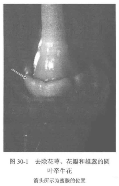
For more product details, please visit Aladdin Scientific website.