Product Manager
Sandra Forbes
Microparticles have exhibited extensive utility across various fields including medicine, biochemistry, colloid chemistry, and aerosol research. Their applications encompass chromatographic separation media, serving as supports for immobilized enzymes, and functioning as spacers in liquid crystal displays. Fluorescently tagged microparticles are valuable as benchmarks in flow cytometry, confocal laser scanning microscopy, and light scattering instruments. Furthermore, they have been employed in environmental science, specifically as tracers in flow measurements of gases and liquids through techniques like laser Doppler anemometry (LDA), particle dynamic analysis (PDA), and particle image velocimetry (PIV).
Melamine Resin Particles
We introduce a novel range of monodisperse polymer microspheres (Figure 1) crafted from melamine resin (MF). These microspheres are synthesized through acid-catalyzed hydrothermal polycondensation of methylol melamine at temperatures ranging from 70-100 °C, eliminating the need for surfactants. By precisely modulating the pH level, concentration of methylol melamine, and reaction temperature, we can achieve a one-pot synthesis of monodisperse particles with a predictable size range of 0.5-15 mm. Thanks to their exceptional physical and chemical properties, melamine resin particles provide numerous advantages compared to traditional polymer particles.
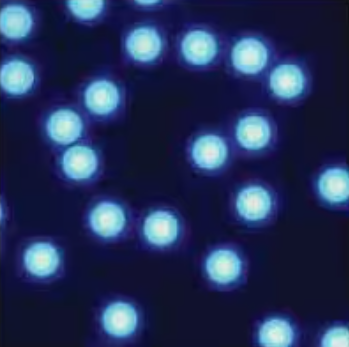
Figure 1. Fluorescence microscopic image of 10 mm melamine resin particles labeled with 7-amino-4-methylcoumarin (reproduced with permission of Microparticles GmbH).
Physical and Chemical Properties of Melamine Resin Particles
·Density: 1.51 g/cm3
·Refractive Index: 1.68
·Excellent monodispersity (C.V. <3%) and highly uniform spherical shape
·Hydrophilic surface
·High crosslinking density
·High thermostability up to 300 °C
·Superior mechanical strength
·Stable and insoluble in acids and bases
·Extremely high stability in organic solvents, no swelling or shrinking upon contact with organic solvents
·Outstanding long-term stability in dispersions; no additives or stabilizers required
·Aqueous suspensions are stable to repeated freeze-thaw cycles
·Particles can be dried directly from their aqueous dispersions
·Free flowing powders of dried particles can be redispersed in any dispersing agent without agglomeration
The unmodified MF particles possess a hydrophilic and charged surface, attributed to the high concentration of polar triazine-amino and -imino groups. These surface functional groups, including methylol and amino groups, facilitate the covalent bonding of other ligands. For specialized applications, MF particles can be modified by the incorporation of additional functionalities, such as carboxyl groups, thereby enhancing the potential for surface derivatization, including chromophore or fluorophore labeling. Melamine resin microspheres are offered as either white particles or those with internally embedded fluorescent labels. Both types of particles are available with either an unmodified surface or one that has been carboxylated.
Fluorescently Labeled and Carboxylate-Modified Melamine Microparticles
Fluorescent melamine resin microspheres can be customized with variations in size, type of fluorochrome, and surface functional groups. Common dyes employed in the creation of MF fluorescent particles include:
·FITC, green fluorescence (λEx = 506 nm, λEm = 529 nm)
·Rhodamine B, orange fluorescence (λEx = 560 nm, λEm = 584 nm)
·Nile Blue A, red fluorescence (λEx = 636 nm, λEm = 686 nm).
The fluorescent MF particles (Figure 2) are distinguished by their narrow size distribution, intense color, and bright fluorescence. These particles are uniformly stained throughout their volume, ensuring no leakage of internally incorporated fluorochromes. They exhibit high stability in organic solvents, comparable to that of white MF particles. Additionally, fluorescent MF beads are available with carboxylate-modified surfaces, featuring a high density of functional groups (>0.1 mmole per gram of resin).
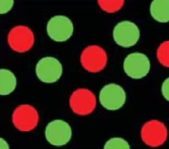
Figure 2. Fluorescence microscopy image of FITC-labeled MF particles (shown as green) and Rhodamine B-labeled MF particles (shown as red).
Fluorescent Nanobeads (Nanoparticles)
In recent years, our understanding of nanoscale systems has seen rapid advancements, leading to the emergence of a new generation of high-tech products and processes. Nanotechnology holds the potential to revolutionize a diverse array of applications, spanning chemicals, electronics, sensors, and advanced materials. Notably, nanoparticles have even been employed as carriers for DNA in drug delivery systems.
A pivotal aspect of nanotechnology involves the development of fluorescent nanoparticles, which could find applications in optical data storage, as well as in the fields of biochemistry, bioanalytics, and medicine. Traditional fluorescent imaging techniques primarily rely on dye markers, which suffer from limited photostability and light emission per molecule. However, nanoparticles offer a solution by providing more intense and stable fluorescent signals. Fluorescently labeled nanoparticles have proven successful in various immunoassay applications. By utilizing very small particles, some of the limitations encountered in latex agglutination assays can be overcome, as they reduce background absorbance and enhance colloidal stability.
Base Materials
Fluorescent nanobeads (or nanoparticles) can be synthesized using various polymers, each imparting specific benefits.
Polyacrylonitrile (PAN) nanoparticles are particularly well-suited for Förster Resonance Energy Transfer (FRET) applications. When labeled, they exhibit high fluorescence and are extremely minute, with diameters less than 30 nm. PAN contains a low concentration of interfering substances and can be obtained with carboxylated or streptavidin-modified surfaces. All particles are provided as a buffered aqueous suspension at 0.5% (w/w) concentration in 10 mM MES buffer, adjusted to pH 7.
PD is an innovative polymer that offers properties akin to polystyrene but boasts significantly lower oxygen permeability, leading to enhanced photostability for most dyes. The particle size is approximately 40 nm, and the surface can be carboxylated or modified in other ways. These particles are also supplied in a buffered aqueous suspension at 0.5% (w/w) concentration in 10 mM MES buffer, adjusted to pH 7.
Custom Particles
Customized microparticles and nanobeads featuring specific modifications, such as streptavidin, are also offered. For inquiries about these tailored products, please contact your local office or sales representative.
Aladdinsci: https://www.aladdinsci.com