Licia Miller Product Manager
The detection of cell surface proteins mainly targets antigens on the cell membrane. Antibodies can directly bind to antigens on the cell surface, so fixation and permeabilization treatment are usually not required. The experimental operation is relatively simple, and the main focus is on the specificity of the antibody and the optimization of its concentration.
However, the detection of nuclear proteins such as FoxP3 and transcription factors requires special fixation and permeabilization steps. These proteins are located in the cell nucleus, and antibodies cannot directly penetrate the cell membrane and nuclear membrane to bind to them. Therefore, the cell structure must be fixed by fixation treatment, and the cell membrane and nuclear membrane must be made permeable using a permeabilization buffer to allow the antibody to enter the cell nucleus and bind to the target protein. In addition, transcription factor detection also requires optimization of fixation and permeabilization conditions to reduce nonspecific background staining and ensure the accuracy and reliability of experimental results.
This article mainly introduces the commonly used experimental operations of cell fixation and permeabilization when performing flow cytometric staining of transcription factors.
1. Preparation before the experiment
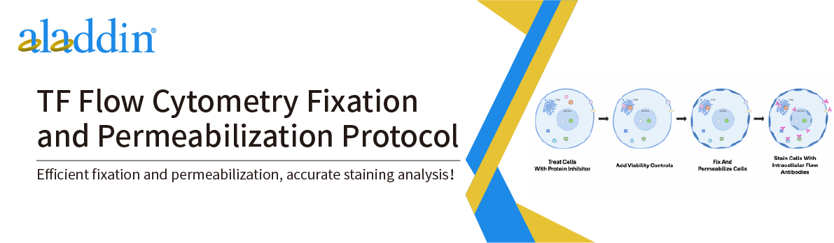
For adherent cells or tissue samples, they must first be dissociated into a single cell suspension.
If processing whole blood samples, lyse red blood cells using Red Blood Cell Lysis Buffer (R478427) prior to fixation and separate cells by centrifugation followed by washing.
If a fixable live or dead cell identification dye is used, it should be added prior to fixation and the excess dye removed before continuing with the fixation step.
Antibodies targeting cell surface markers (e.g., CD4+ markers) can be added prior to fixation and remain bound to the target antigen during fixation. If necessary, a wash can be performed prior to fixation.
Centrifugation conditions need to be adjusted according to cell type and sample volume, and 150-300℃ is generally recommended. Centrifuge at 4℃ for 1-5 min.
2. Fixation and Permeabilization
1. Centrifuge to obtain cell pellet and remove supernatant.
2. Resuspend cells in 1 mL 4% paraformaldehyde and mix gently to disperse cell clumps and avoid cross-linking.
3. Incubate at room temperature (20-25℃) in the dark for 10-15 minutes.
4. Wash cells twice with 1× PBS.
5. Add 100 µL of 1× PBS containing 0.1% Triton X-100 per 10⁶ cells and incubate at room temperature for 10-15 minutes. Centrifuge and discard supernatant.
6. Wash cells twice with 1× PBST containing 0.05%-0.1% Tween-20.
7. Block cells with blocking solution (such as normal goat serum or BSA) and incubate at room temperature for 30-60 minutes.
3. Immunofluorescence staining
1. Cell Count: Count the cells using a hemocytometer or other method.
2. Dispense the cells into test tubes or wells according to experimental requirements. It is recommended to use 5×10⁵ to 1×10⁶ cells for each test.
3. Dilute the antibody according to the recommended dilution concentration of the antibody product with 100 µL 1× PBST, then aspirate the antibody dilution solution to resuspend the cells.
4. Incubate at room temperature (20-25℃) in the dark for 1 hour.
5. Wash the cells by centrifugation with sufficient 1× PBST, discard the supernatant, and repeat the washing step twice.
6. If using a fluorescein-conjugated primary antibody, resuspend the cells in 200-500 µL 1 × PBST directly after washing and analyze by flow cytometry. If using an unconjugated primary antibody, proceed to the next step.
7. Dilute the secondary antibody conjugated to fluorescein at the recommended dilution concentration in 1 × PBST and resuspend the cells.
8. Incubate at room temperature (20-25℃) in the dark for 30 minutes.
9. Wash the cells by centrifugation using 1× PBST, discard the supernatant, and repeat the washing step twice.
10. Resuspend the cells in 200-500 µL of 1 × PBST for flow cytometry analysis.
For more product details, please visit Aladdin Scientific website.