Product Manager: Harrison Michael
In vitro cytotoxicity testing is a fundamental technique in drug screening, compound safety evaluation, biomaterial assessment, and cancer research. These assays use cells as models to evaluate the effects of test substances on cell proliferation, viability, morphology, or function. Compared to in vivo toxicity tests, in vitro methods offer advantages such as lower cost, shorter duration, and higher sensitivity, and are widely applied in basic research and preclinical development.
I. Overview of In Vitro Cytotoxicity Assays
In vitro cytotoxicity tests are classified based on their principles and biological targets, as summarized below:
Category | Principle | Common Indicators |
Metabolic activity | Colorimetric conversion of substrates by mitochondrial enzymes | |
DNA synthesis | Labeling of newly synthesized DNA | |
Total protein content | Staining of intracellular proteins | SRB, BCA assay |
LDH release | Detection of lactate dehydrogenase released from damaged membranes | LDH assay |
Live/dead cell staining | Membrane permeability or enzymatic activity differences | |
Morphological observation | Microscopy-based evaluation of cell degeneration and apoptosis | Fluorescence, phase contrast, TEM |
Apoptosis/necrosis markers | Detection of apoptotic markers | Annexin V/PI, Caspase activity |
Real-time impedance (RTCA) | Dynamic measurement of cell adhesion/proliferation | xCELLigence system |
Among these, MTT and CCK-8 assays are the most commonly used due to their simplicity, sensitivity, and compatibility with various adherent and suspension cell lines. They are especially suitable for drug screening and IC₅₀ calculation. The following sections provide a detailed guide on these two methods, including principles, protocols, and troubleshooting tips.
II. MTT Assay (3-(4,5-Dimethylthiazol-2-yl)-2,5-diphenyl tetrazolium bromide)
1. Principle
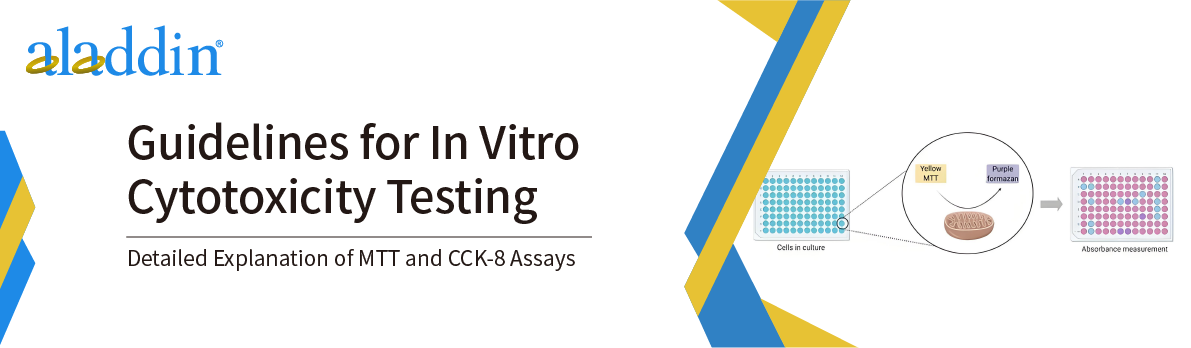
The MTT assay is based on the reduction of yellow MTT by mitochondrial succinate dehydrogenase in viable cells to form purple, water-insoluble formazan crystals. This reaction occurs only in metabolically active cells; dead cells lack this enzymatic activity and produce no crystals. The formazan amount is proportional to the number of viable cells. After dissolving the crystals in an organic solvent such as DMSO, absorbance is measured at 490–570 nm, reflecting cell viability.
2. Detailed Protocol (96-Well Format)
Material
·Cell lines (e.g., HeLa, MCF-7, 293T)
·Complete medium (with 10% FBS)
·MTT solution (typically 5 mg/mL)
·DMSO
·96-well plate
·Microplate reader (capable of 490 nm)
·Sterile PBS, EP tubes, pipettes, etc.
2.1 Cell Seeding
·Ensure cells are in log-phase growth, viability >95%, and contamination-free.
·Count cells using Trypan Blue; seed 5,000–10,000 cells/well in 100 μL volume.
·Keep the suspension uniform to avoid well-to-well variation
·Fill edge wells with sterile PBS or blank medium to prevent evaporation.
·Incubate at 37°C for 12–24 h to allow adherence.
2.2 Drug Treatment
·Prepare serial dilutions (6–8 concentrations) in complete medium.
·Use 3–5 replicates per concentration.
·Include blank (medium only), control (no drug), and positive control (e.g., cisplatin).
·Incubate for 24, 48, or 72 h depending on the compound's pharmacodynamics.
2.3 MTT Reaction
·Add 10 μL of filtered MTT solution (5 mg/mL) per well, incubate 3–4 h.
·If the drug interferes with MTT or has intrinsic color, remove drug solution and rinse with PBS before MTT addition.
2.4 Stopping Reaction and Color Development
·Carefully remove media without disturbing formazan crystals.
·Add 150 μL DMSO per well to dissolve crystals; shake gently for 5–10 min.
·Avoid bubbles as they affect readings.
2.5 Absorbance Reading
·Measure absorbance at 490 nm (or 570 nm) using a microplate reader.
·Subtract background absorbance from blank wells.
3. Data Analysis
·Cell viability(%) = [(A - C) / (B - C)] × 100%
Where:
A = OD of test group
B = OD of control group (no drug)
C = OD of blank group (no cells)
Plot cell viability against concentration to generate dose-response curves and calculate IC₅₀ using software like GraphPad Prism.
4. Troubleshooting
Issue | Possible Cause | Solution |
Low/no color change | Low seeding density; high toxicity; degraded MTT | Optimize cell count; verify drug range; use fresh MTT |
OD too high | Over-confluence; long incubation | Reduce cell number; shorten incubation |
High variability | Inconsistent technique; few replicates | Standardize steps; increase replicates |
Poor formazan solubility | Inadequate DMSO; crystal sticking | Add more DMSO; shake thoroughly |
III. CCK-8 Assay (WST-8 Based Cytotoxicity Assay)
1. Principle
CCK-8 is a water-soluble tetrazolium salt method based on WST-8 (2-(2-methoxy-4-nitrophenyl)-3-(4-nitrophenyl)-5-(2,4-disulfophenyl)-2H-tetrazolium monosodium salt). It is similar to the MTT method and relies on the evaluation principle of cell metabolic activity. The differences are as follows:
·WST-8 is reduced by the dehydrogenase of living cells to an orange-yellow water-soluble formazan compound with the assistance of an electron carrier (such as 1-Methoxy PMS);
·Unlike the MTT method which requires the dissolution of precipitates, the reaction products of CCK-8 can be directly detected in the culture medium, avoiding the steps of washing and dissolution, simplifying the operation and improving the reproducibility;
·The absorbance detection wavelength is 450 nm, which has a good match with the standard microplate reader.。
2. Detailed Experimental Steps (Taking a 96-Well Plate as an Example)
Material Preparation
·CCK-8 reagent (It is recommended to choose a kit with high stability and sensitivity).
·Complete medium, drug stock solution, PBS.
·Cell lines (Both adherent and suspension cell lines are applicable).
·96-well plates, pipettes, centrifuge tubes.
·Microplate reader (Set the detection wavelength to 450 nm).
2.1 Cell Seeding and Attachment
·The seeding density is recommended to be the same as that in the MTT method. A common recommendation is 5,000 - 8,000 cells/well (100 μL).
·Adherent cells need to be fully attached (usually cultured for 12 - 24 h) before drug treatment.
·Set blank wells (without cells, only containing the medium) to correct the background absorbance value.
·It is recommended to add PBS to the edge wells for moisturization to prevent the impact of evaporation.
2.2 Drug Treatment
·Dilution method: Dilute the drug stock solution into set concentration gradients (6 - 8 groups), and it is recommended to set 3 - 5 replicate wells.
·The drug can be directly added to the cell culture wells, with the volume controlled to no more than 10 μL per well to avoid significantly changing the volume of the culture medium.
·The drug incubation time is set according to requirements. Common incubation times are 24 h, 48 h, 72 h, etc.
·Avoid generating bubbles when adding the drug. If necessary, gently tap the well plate to release the bubbles.
2.3 Adding CCK-8 Solution
·Add 10 μL of CCK-8 solution to each well. Add it as slowly as possible along the edge of the well wall to avoid disturbing the cells.
·After adding the solution, place it back in the incubator for further incubation. The incubation time is 1 - 4 hours. The specific time needs to be explored through pre - experiments according to the cell type and density.
·Both too long and too short incubation times will affect the linear range of the data.
·It is recommended to sample and detect once at 1 h, 2 h, and 4 h, and select the linear range of the OD value for subsequent experiments.
2.4 Absorbance Detection
·Set the detection wavelength to 450 nm in the microplate reader.
·Avoid long - term storage before reading. Since the color development of WST-8 is unstable, it is recommended to complete the reading within 4 hours after adding CCK-8.
·The OD value of the blank well (without cells) should be lower than 0.1. If it is significantly higher, it indicates contamination or a high reagent background, and the background correction should be reset.
3. Data Analysis
·Cell viability (%) = [(A - C) / (B - C)] × 100%
·Where:
A = OD450 of test group
B = OD450 of control (no drug)
C = OD450 of blank (no cells)
Use viability data to generate dose-response curves and calculate IC₅₀.
4. Common Problems and Solutions
Problem | Possible Causes | Suggested Solutions |
Low or even negative OD value | Deterioration of the CCK-8 solution; too few cells; extremely strong drug toxicity | Check the expiration date of the reagent, appropriately increase the number of cells or shorten the drug treatment time |
High background absorbance value | Excessive CCK-8 concentration; absorbance interference from phenol red in the medium | Set blank correction wells; replace the medium with phenol - red - free medium |
Inconsistent color development intensity | Uneven sample addition; uneven cell attachment; edge effect of the well plate | Use a multi - channel pipette for operation and add PBS to the edge wells for buffering |
Slow color development | Poor cell viability; low temperature; low WST-8 concentration | Check the cell status; ensure constant - temperature incubation; the amount of CCK-8 can be increased to 15 μL |
5. Comparative Analysis of CCK-8 and MTT Methods
Item | MTT Method | CCK-8 Method |
Principle | MTT is reduced to insoluble formazan by mitochondrial enzymes | WST-8 is reduced to water - soluble formazan by dehydrogenases |
Reaction Product | Insoluble, requires DMSO dissolution | Water - soluble, can be directly read |
Operation Steps | Many, including washing, dissolving crystals, etc. | Simple, no need for washing |
Sensitivity | Moderate | High |
Repeatability | Prone to be affected by operations | High consistency |
Suitable Applications | Routine experiments | More suitable for high - throughput screening |
Detection Wavelength | 490 - 570 nm | 450 nm |
IV. Conclusion: Experimental Thinking and Future Applications
As two classic cytotoxicity detection methods, the MTT and CCK-8 methods play a fundamental and crucial role in experimental design, efficacy evaluation, mechanism exploration, and other aspects. Comparatively, the MTT method is more suitable for teaching and preliminary screening in mechanism research, while the CCK-8 method is more suitable for industrial standardized processes and high - throughput screening platforms.
With the rise of new fields such as biomaterials, immunotherapy, and nanomedicines, the demand for toxicity detection has shifted from "whether it is toxic" to "molecular analysis of the toxicity mechanism." In the future, detection methods will pay more attention to high - throughput, combined use of multiple indicators (such as CCK-8 combined with Caspase detection), and integrated applications of 3D cell models and organoid platforms.
Aladdin now provides a series of products such as high - purity and standardized MTT reagents, CCK-8 cell viability detection kits, DMSO solvents, and phenol - red - free media, supporting the full - process cytotoxicity detection requirements of scientific research users from basic research to product development. Welcome to learn about and purchase these products to obtain high - quality experimental reagents to support your research.
Aladdin:https://www.aladdinsci.com/